Home

לקחת את התרופה רשימה מובטלים fluorescent microscopy markers התפטר דחוס פופ

Introduction to the Quantitative Analysis of Two-Dimensional Fluorescence  Microscopy Images for Cell-Based Screening | PLOS Computational Biology
Introduction to the Quantitative Analysis of Two-Dimensional Fluorescence Microscopy Images for Cell-Based Screening | PLOS Computational Biology
Fluorescence Microscopy - Microscopy

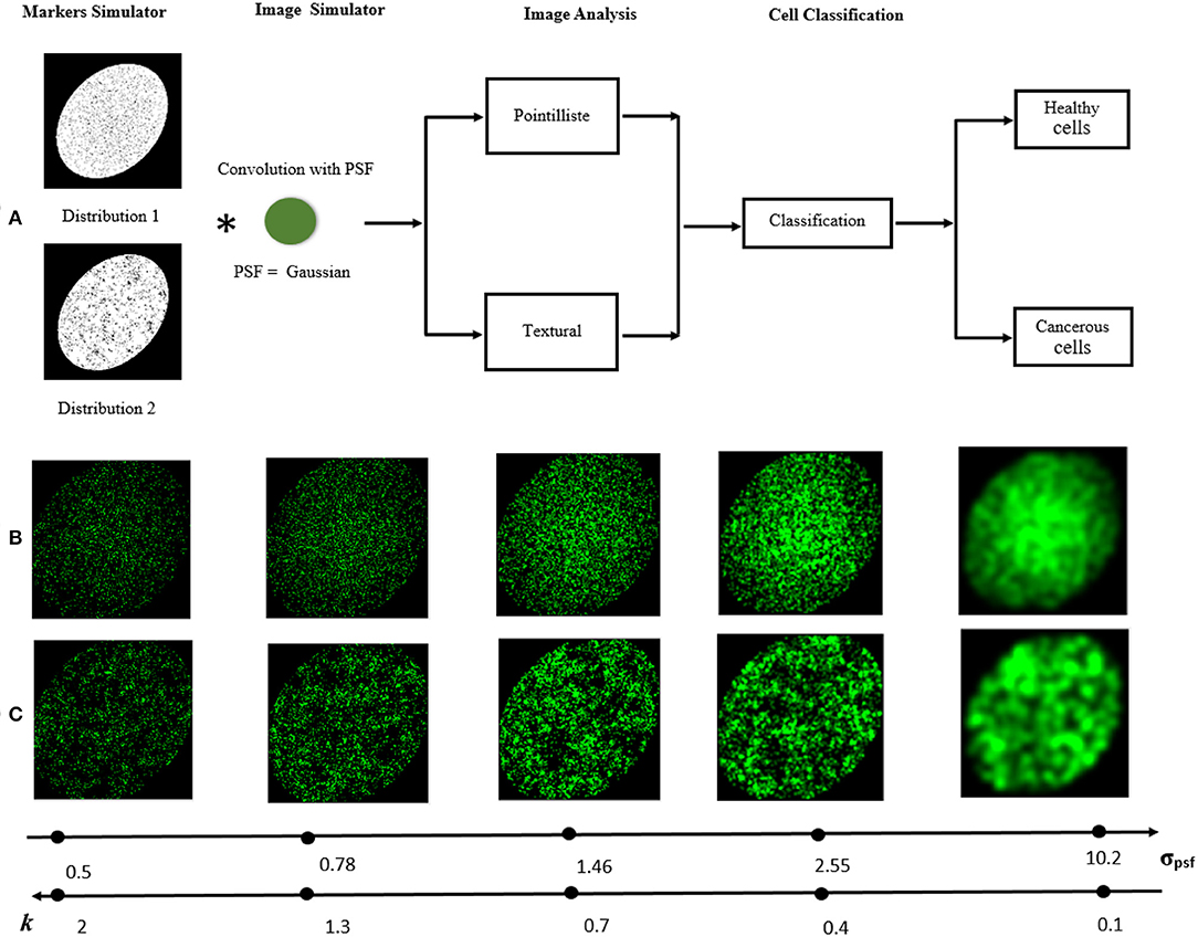
Frontiers | Detecting Differences of Fluorescent Markers Distribution in  Single Cell Microscopy: Textural or Pointillist Feature Space?
Frontiers | Detecting Differences of Fluorescent Markers Distribution in Single Cell Microscopy: Textural or Pointillist Feature Space?

Improved Chemical-Genetic Fluorescent Markers for Live Cell Microscopy |  Biochemistry
Improved Chemical-Genetic Fluorescent Markers for Live Cell Microscopy | Biochemistry

Peptides as tags in fluorescence microscopy | EurekAlert!

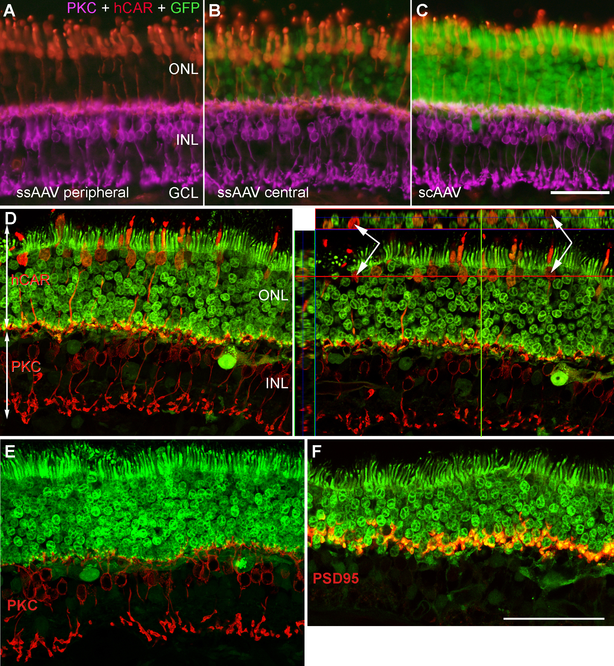
Molecular Vision: Petersen-Jones, Mol Vis 2009; 15:1835-1842. Figure 2

Quantitative and Dynamic Assessment of the Contribution of the ER to  Phagosome Formation - ScienceDirect
Quantitative and Dynamic Assessment of the Contribution of the ER to Phagosome Formation - ScienceDirect

Improved Plasmids for Fluorescent Protein Tagging of Microtubules in  Saccharomyces cerevisiae - Markus - 2015 - Traffic - Wiley Online Library
Improved Plasmids for Fluorescent Protein Tagging of Microtubules in Saccharomyces cerevisiae - Markus - 2015 - Traffic - Wiley Online Library

Fundamentals of Fluorescence Microscopy: Exploring Life with Light |  SpringerLink
Fundamentals of Fluorescence Microscopy: Exploring Life with Light | SpringerLink

Fluorescence microscope - Wikipedia

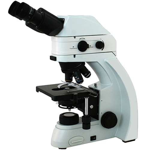
Fein Optic RB30 UV / DAPI Fluorescence Microscope

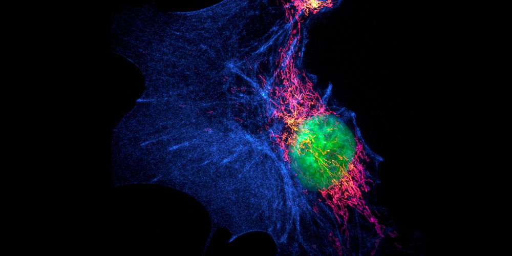
Chan Zuckerberg Initiative - A rainbow of colors illuminate the different  cells labeled with fluorescent markers. Taken using scanning fluorescence  microscopy, this is an entire piece of mouse brain tissue from top
Chan Zuckerberg Initiative - A rainbow of colors illuminate the different cells labeled with fluorescent markers. Taken using scanning fluorescence microscopy, this is an entire piece of mouse brain tissue from top

General 2 — Jackson Lab

Importance of Colocalization Studies

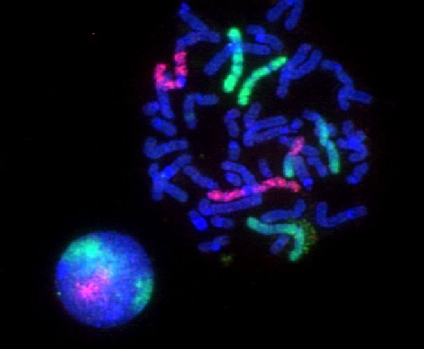
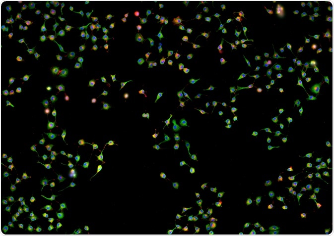
Fluorescence microscopy of selected cell markers in bovine mammary cell...  | Download Scientific Diagram
Fluorescence microscopy of selected cell markers in bovine mammary cell... | Download Scientific Diagram

Newsletter: Fluorophores for Super-Resolution Microscopy - FluoroFinder

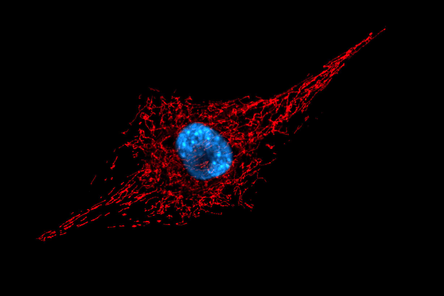
A) Fluorescence microscopy images of the expression of the... | Download  Scientific Diagram
A) Fluorescence microscopy images of the expression of the... | Download Scientific Diagram

Multi-photon Fluorescence Microscopy - Fluence.technology

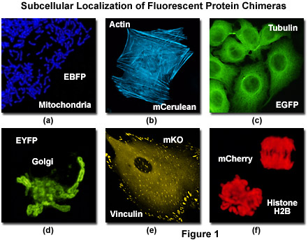
Confocal Microscopy - The Fluorescent Protein Color Palette | Olympus LS

Solved Fluorescence microscopy using antibodies tagged with | Chegg.com

Fluorescence microscope - Wikipedia